NIF Human Imaging Facility
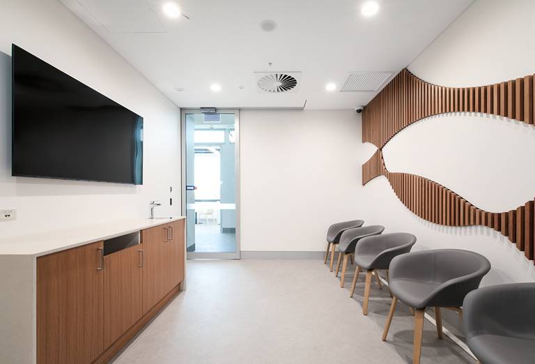
The WA NIF node Human Imaging Facility provides in vivo human imaging capabilities, welcoming two state-of-the-art research-dedicated scanners: a digital PET-CT and a 3T MRI system.
Our human imaging systems
These new scanners are the only human imaging systems in WA fully dedicated to research, facilitating high-end in vivo human imaging for the state. They are co-located with our preclinical instruments, offering unique advantages to human participants and researchers, streamlining workflows, and opening the path towards multimodal and co-registered imaging.

3T Magnetic Resonance Imaging (MRI)
Magnetic resonance imaging (MRI) uses applied magnetic fields to non-invasively (and non-destructively) image samples with non-ionising radiation (unlike X-ray based techniques). MRI can rapidly provide excellent in vivo soft-tissue image contrast for qualitative analyses, 3D images for quantitative volumetric measurements, and access to quantitative parameter maps (such MR relaxation, diffusion, flow) related to underlying tissue structure, function, or metabolism. Rapid imaging techniques can also be used to study dynamic processes, such as the cardiac cycle.
Using ultra-short echo time techniques we can provide CT-like images of solid tissues like bone, to completement MRI’s exquisite soft-tissue contrast within the same imaging session.
Unique to the MRI research landscape in WA, the WA NIF node has MR Elastography (MRE) equipment to non-invasively provide estimates of tissue stiffness. MRE has quickly become an invaluable marker of liver fibrosis and nonalcoholic steatohepatitis (NASH), being an ideal imaging biomarker for clinical trials into new drugs to treat these conditions.

Human Computed Tomography (CT)
.jpeg?h=540&iar=0&w=720&hash=803E82244A0C5D24FBD709F36CC13A86)
Positron Emission Tomography (PET)
FAQ
-
Can we do stand-alone CT scans on the PET/CT scanner?Yes, we have a Siemens Edge 128-slice CT system which can do diagnostic CT scans. We also have a power injector for injecting CT contrast agent (Medrad Stellant FLEX from Bayer).
-
What PET radiotracers are available at WA NIF?Get in touch with us to discuss tracer availability and possibilities.
-
Is your PET/CT scanner ARTnet accredited?Yes. We have a general F-18,Ga-68 and Zr-89 ARTnet accreditation. We can also perform trial-specific phantoms or other radiotracers on request.
-
How do we get started to use WA NIF for our study?Get in touch with us to get started. We can provide you with a quote and advice on further requirements (e.g. ethics and governance related to your institution).
-
Do you have a Medicare licence?No, WA NIF does not currently have a Medicare licence for the PET/CT or MRI.
-
How much does it cost to use your facility?Please get in touch with us to get a custom quote. The quote will depend on multiple factors such as scan length, protocol and uptake time, among others. Scanning fees will also depend if its an investigator-led, academic study or a pharmaceutical-sponsored trial. We can also help you with your study budget for an funding application.
-
Can we use your facility for diagnostic work?We only do scans as part of research studies at WA NIF.
-
How do we book in study participants for WA NIF?We have a booking system. Get in touch to get started.
-
Do you have offer parking for study participants at WA NIF?Parking is available on the QEII Hospital Campus. We recommend parking in the DD block carpark.
-
Can we collaborate with your facility on an upcoming study?Yes. We have research fellows specialised in PET radiopharmaceuticals, PET/CT and MRI who are very keen to collaborate and help you with any scientific questions you may have.

Location and contact
Email: [email protected]
Address: WA National Imaging Facility
Level 3, Harry Perkins Institute of Medical Research
QEII Medical Centre campus
Nedlands WA 6009
Our partners
Major partners


Major supporter Host institution


Supporters


